


This disease is poorly understood, although several thousand observations with diagnosis and subsequent treatment have been described.
The multitude of diversity and non-specificity of the clinical picture of small pelvic varicose veins leads to serious errors in diagnosis, which in the future affect the consequences.
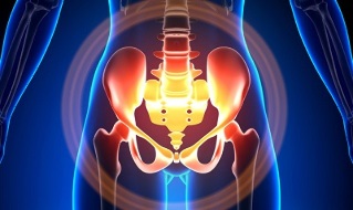
Characteristics of small pelvic varicose veins
The pelvic veins are several times larger than the arteries, which leads to their larger capacity. This is due to the phylogeny of the vascular system of the pelvic area. The pelvic veins are very adaptable and possibly prone to remodeling, which helps to create a dense interconnected network.The speed and direction of blood flow are regulated by valves, which are controlled by complex suction mechanisms. The valves balance the pressure in different parts of the venous network.
When the valves stop performing their functions, blood stasis develops, which leads to vascular pathology and the formation of varicose veins. The uniqueness of the pelvic veins lies in the fact that the wide ligaments of the uterus, which maintain the lumen of the vessel, can narrow it, causing pathology.
Causes of occurrence
Abnormal dilation of the venous pelvis may be due to the following reasons:
- Blood flow disorder?
- Elimination of the vein trunk?
- Compression of logs from a distorted position of the uterus, for example, in retrospect.
- ovarian valve insufficiency (congenital or acquired);
- Obstructive post-venous syndrome?
- Connective tissue pathology?
- Arteriovenous angioplasia?
- Prolonged sitting, hard physical work
- Varicose veins of the lower extremities?
- Pregnancy (3 or more) and childbirth (2 or more) Diseases of the female genital area (chronic fallopian tube infection, ovarian tumors, uterine fibroids and genital endometriosis).
- Adhesion of the pelvic organs?
- Obesity.
Classification by degree of disease
The following degrees are distinguished by the size of the dilated vein:
- up to 0. 5 cm, "opener" of boats.
- 0, 6-1 cm;
- more than 1 cm.
Variations in the course of the disease
- varicose veins of the perineum and atrium of the vagina.
- small pelvic venous congestion syndrome.
Symptoms
- The most common - frequent pain in the lower abdomen, the perineum after long static and dynamic overexertion. The pain intensifies in the second phase of the cycle, after hypothermia, fatigue, stress, exacerbations of various diseases.
- Feeling "out of place", pain during and after sex. Dysmenorrhea - menstrual abnormalities, including pain.
- Excretion, more than normal, of the glands of the genital system.
- Blood stasis leads to infertility, miscarriage, abortion.
- Violation of urination due to the expansion of the veins of the bladder.
Diagnostics
Diagnosis of the disease only with complaints is successful in only 10% of cases.
The palpation of the inner walls of the pelvis makes it possible to feel the elongated seals and the venous nodes. When observed in mirrors, cyanosis of the vaginal mucosa is visible.
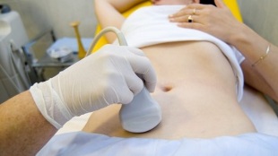
The selection procedure is an ultrasound examination with color Doppler mapping, which allows the detection not only of varicose veins of the ovaries, but also of venous thrombosis, post-thrombophlebotic obstructions. The ultrasound shows a tortoise, "like a worm", structures without signal reflection, located on the lateral surface of the uterus.
The Doppler effect is based on "hue" in blue and red, venous and arterial blood flow, respectively.
The device for ultrasound examination with the help of a special program recognizes the movement of blood from the sensor and in the other direction, calculates the speed of blood flow and the type of vessel.
But the exact definition of the vein or artery remains with the doctor. The Doppler method works in almost all cases, exceptions to the rules are dictated by our body, as the blood flowing from the heart is not always arterial and vice versa.
Thus, the diagnostic ultrasound doctor looks at this arterial or venous vessel, its size, the rate of blood flow in it, and many indicators that do not need an ordinary person, but play an important role in the diagnosis. This is why transdermal and transvaginal sensors are used.
In 5. 7% of cases, the disease is randomly identified during screening. Normally, the diameter of the ovarian vein is 0. 4 cm.
CT and MRI are very accurate. With these methods, it is possible to detect the accumulation of varicose veins in the ligaments of the uterus, ovaries and around these organs. It is possible to determine the concomitant pathology.A very reliable method is venography research.
The contrast takes place at the height of the Valsalva test, versus blood flow. This allows you to see exactly the valve failure.
Left retenogenoscopy, renal venography, hyper-selective venobarioscopy, and venobariography on both sides are also used. These methods make it possible to identify hemodynamic and anatomical changes in the renal veins and the sites where the gonadal veins flow. Hyperselective venobaryoscopy is performed by catheterization of the gonadal veins through the contralateral femoral or subclavian vein, followed by contrast injection.Most of the blood from the varicose veins of the urinary tract is excreted through the ovarian vein. But in hypertensive conditions, it occurs through the exogenous veins of the uterus to the internal iliac vein. The network of veins through which outflow can occur includes the sacrum and the bladder.
On the left side of the venobaricography, there are 3 stages of venous stasis in the ultraviolet grid of the left ovary:
- There is no outflow from the left ovarian plexus or an extra short path follows.
- There is an extra long route.
- Two additional outflow paths are visible or one additional and auxiliary.
In stages 2 and 3, varicose veins of the right ovary form.
Laparoscopy is used for differential diagnosis. The abnormally helical veins are located in the area of the ovaries, in the direction of the round and wide ligaments. They look like large cyanotic bands with a thin and tense wall.
The complexity of the diagnosis lies in the fact that the disease often hides behind signs of inflammatory process, differs in clinical manifestations, disguised as endometriosis, prolapse of internal organs, postoperative neuropathies and many exogenous diseases.
Treatment
The main goal of treatment is to remove the regression in the veins. In the early stages of the disease, conservative treatment is used. In the later stages of the disease, surgery is the treatment of choice.
Conservative treatment

It consists in the normalization of venous tone, in the improvement of hemodynamics and trophic processes.
Symptomatic treatment for individual symptoms. Non-steroidal anti-inflammatory drugs for pain, bleeding - hemostatic therapy.
The main drugs in conservative treatment are ventonic drugs and antiplatelet agents.
Phlebotonics - improve vascular wall tone and increase blood flow. With this disease, it is best to consult a gynecologist about certain medications.
Physiotherapy is an important method.
Surgical treatment
- Varicose veins resection.
- Gonado Hippodrome.
- Hardening on laparoscopy.
- Obstruction of the ovarian veins using endovascular X-ray methods
Folk remedies
Because the main factor in the onset of the disease is the weakness of the valve device, all these folk remedies used for varicose veins of the lower extremities are also used for this pathology.
The most commonly used are: common hazelnut, hops, nettles, chestnuts, dandelion root, kombucha, willow, oak, St. John's wort, string, pollen and many other plants.
The following are effective: bath treatment with oak, chestnut, willow, chamomile, pharmacy, cayenne herbs, St. John's word, string.
Prevention
- The first thing you should do if you have any complaints, predictions or illnesses listed above is to contact your gynecologist.
- It is necessary to normalize the work regime and rest, try not to stay upright for a long time, physical excessive pressure.
- Do "pedal", "birch", "scissor feet" prevention exercises
- Stay on a diet: eat foods rich in vitamins E, P, C, try to eat only white meat, less fatty meat, replace it with fruits, vegetables, cereals.
- Drink plenty of fluids, but not less than 1. 5 liters per day.
- Get rid of excess weight, bad habits.
- Consult your doctor about the use of compression garments, it will improve blood flow from the lower extremities, thus reducing congestion in the pelvis.
- Avoid baths, saunas, steam baths, hot baths.
In order not to get sick with such a difficult disease to diagnose, you must follow the preventive recommendations mentioned above. Treat your health as the most precious thing in life.
For the slightest suspicious symptoms that you can not get rid of in a few days, you should visit your doctor. He must provide you with specialized help and save you from suffering.